
Many health-conscious individuals invest thousands of dollars in exotic stem cell therapies or unproven longevity elixirs, yet overlook a fundamental biological bottleneck occurring silently within their cells: the epigenetic lock caused by the depletion of a simple endogenous metabolite. This molecule is Alpha-Ketoglutarate (AKG). For decades, AKG was relegated to biochemistry textbooks as a mundane intermediate in the mitochondrial Krebs cycle, responsible solely for generating ATP. However, pioneering research in longevity medicine has unveiled a far more profound role: AKG acts as a master regulator of the epigenome. As endogenous AKG levels plummet with age, DNA demethylating enzymes are starved of their essential co-factor, triggering epigenetic drift, one of the primary hallmarks of aging. Understanding how AKG orchestrates epigenetic reprogramming not only allows us to optimize physical performance but also unlocks a scientifically validated pathway to reverse biological age.
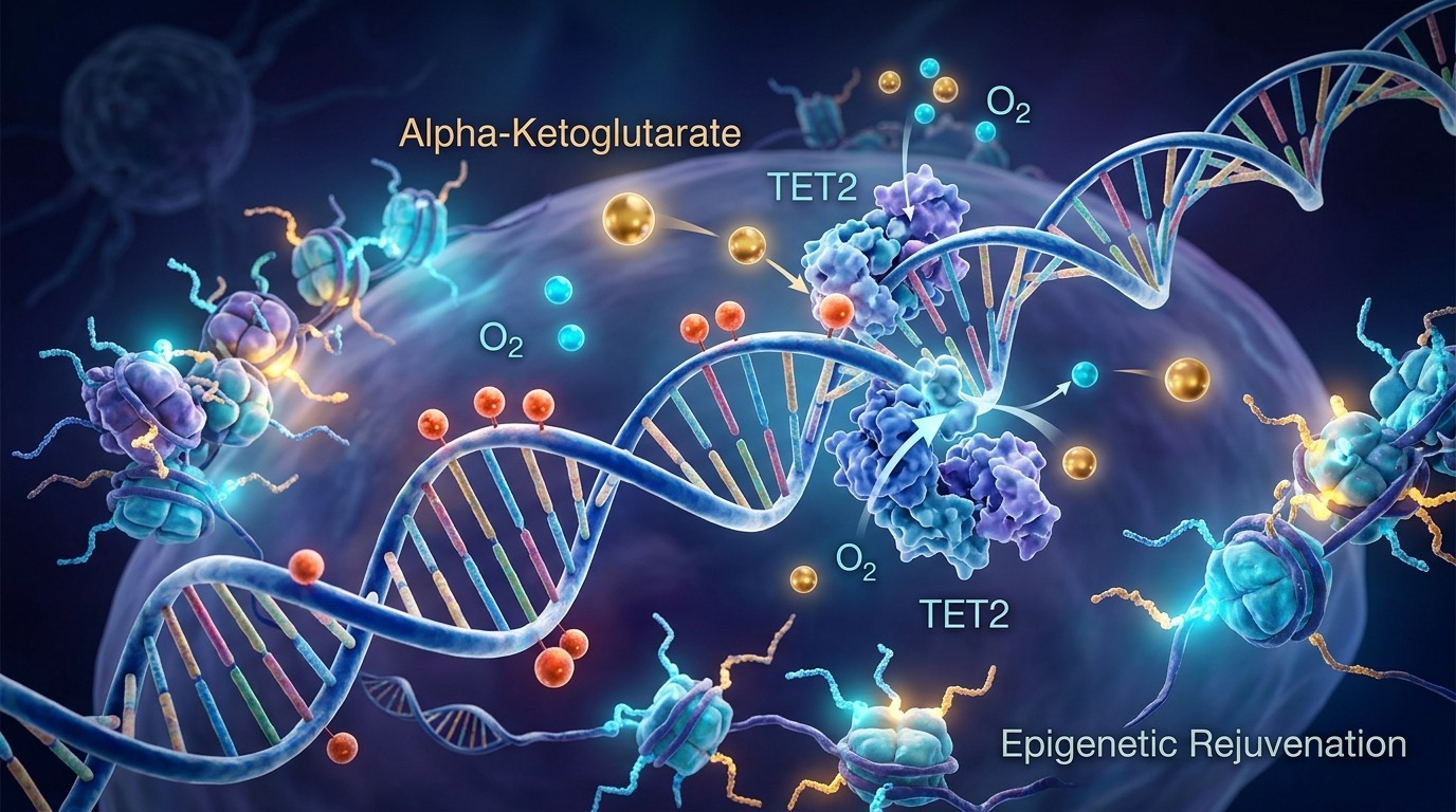
"Imagine our genome as a vast library containing tens of thousands of instruction manuals for cellular operation. Over time, sticky dust particles (methyl groups, -CH3) accumulate on the pages, locking important repair manuals shut and preventing the cell from reading critical survival instructions. The TET enzymes act as diligent librarians, tasked with cleaning and removing these stubborn stains to restore the pages to their pristine, readable state. However, these librarians are completely paralyzed without their specialized cleaning solvent: Alpha-Ketoglutarate (AKG). As we age, the cellular pool of AKG dries up, leaving the epigenetic library cluttered and dysfunctional. Supplementing with AKG replenishes this vital solvent, reactivating the TET enzymes to sweep away the epigenetic dust of aging."
Molecular Pathway Flowchart
Calcium-AKG Supplementation
Cellular Import via SLC13A3
Elevated Intracellular AKG Pool
Activation of TET1/2/3 Enzymes
DNA Demethylation (5mC to 5hmC)
Reactivation of Longevity Genes & NF-kB Inhibition
1. Molecular Mechanisms: The TET-KDM Axis and Epigenetic Reprogramming
To understand how Alpha-Ketoglutarate (AKG) reverses biological age, we must delve into the intricate machinery of epigenetics. While our underlying genetic code remains static, the expression of these genes is dynamic, controlled by methyl groups (-CH3) bound to DNA (which silence genes) and the chemical modifications of histone proteins.
AKG serves as an obligatory co-factor for a superfamily of enzymes known as 2-oxoglutarate-dependent dioxygenases (2-OGDDs). Among these, the Ten-Eleven Translocation (TET1, TET2, TET3) DNA demethylases and Jumonji C domain-containing histone demethylases (KDMs) are paramount.
TET enzymes utilize oxygen and catalytic iron (Fe2+) to oxidize 5-methylcytosine (5mC, the silenced state) into 5-hydroxymethylcytosine (5hmC, the active state), initiating active DNA demethylation. Concurrently, KDMs remove repressive methyl marks from histone tails, relaxing the chromatin architecture to allow transcription factors access to essential longevity genes such as Sirtuins and FOXO3. In the absence of adequate AKG, these epigenetic remodelers are starved, locking the cell into a senescent, hypermethylated state.
2. The Epigenetic-Metabolic Paradox & Comparative Clinical Metrics
A profound biological paradox lies in the spatial compartmentalization of AKG: it is synthesized within the mitochondrial matrix via the tricarboxylic acid (TCA) cycle by Isocitrate Dehydrogenase (IDH), yet its primary epigenetic targets reside within the nucleus. The efflux of AKG across the mitochondrial inner membrane via the dicarboxylate carrier (SLC25A11) is the critical rate-limiting step for epigenetic signaling.
During mitochondrial decay associated with aging, not only does absolute AKG production plummet, but its structural analogs, Succinate and Fumarate, accumulate due to the downregulation of Succinate Dehydrogenase (SDH). Succinate and Fumarate act as potent competitive inhibitors at the AKG-binding pocket of TET and KDM enzymes. Consequently, the intracellular AKG-to-Succinate ratio, rather than absolute AKG concentration, dictates the epigenetic landscape.
| Physiological State | AKG/Succinate Ratio | Epigenetic Landscape | Longevity Gene Expression | Inflammatory Secretome (SASP) |
|---|---|---|---|---|
| Youthful Homeostasis | High (> 5:1) | Active demethylation, open chromatin | Robust expression (SIRT1, FOXO3) | Minimal |
| Chronological Aging | Low (< 1:1) | Hypermethylation, closed chromatin | Suppressed, silenced | High (Elevated IL-6, TNF-alpha) |
| Ca-AKG Intervention | Restored Ratio | Epigenetic reprogramming, 5mC clearance | Reactivated transcriptional pathways | Suppressed, reversed inflammaging |
3. Practical Takeaways: Clinical Protocols and Safety Guidelines
Translating the science of AKG into clinical efficacy requires careful consideration of pharmacokinetics. Administering AKG as a free acid is highly inefficient due to its chemical instability and potential to induce mild metabolic acidosis in the gastric environment.
Instead, Calcium-AKG (Ca-AKG) is the preferred formulation utilized in landmark clinical trials. The calcium bound to AKG slows its release in the gastrointestinal tract, enhancing systemic bioavailability and maintaining stable plasma levels. The landmark human clinical trial using Ca-AKG (the Rejuvant study) demonstrated an average reduction of 8 years in biological age (measured by the DNAmFitAge methylation clock) after 7 months of supplementation.
Clinical Recommendations: - Dosage: 1000mg to 1500mg of Ca-AKG daily for individuals over the age of 40. - Timing: Administer with morning or afternoon meals to maximize absorption alongside dietary micronutrients. - Synergistic Combinations: Co-administer with Vitamin C (which maintains iron in its active Fe2+ state, a critical co-factor for TET enzymes) and AMPK activators such as Berberine to optimize mitochondrial efficiency. - Safety Monitoring: Patients with a history of calcium-oxalate nephrolithiasis should consult their physician and monitor urinary calcium excretion during long-term, high-dose therapy.