
For decades, conventional health messaging has framed insulin merely as a benevolent metabolic janitor whose only duty is sweeping glucose out of the bloodstream. In fundamental cellular biology, however, insulin wields a vastly more powerful identity: The ultimate anabolic hormone.
The ancient evolutionary command embedded within an insulin surge is unmistakable: "Nutrients are abundant; absorb building blocks, store fuel, and accelerate cellular division!"
When systemic insulin levels and its molecular cousin IGF-1 (Insulin-like Growth Factor 1) remain persistently elevated over years due to visceral adiposity and insulin resistance, this physiological growth signal becomes a jammed accelerator pedal, driving premalignant cells into nonstop mitotic division.
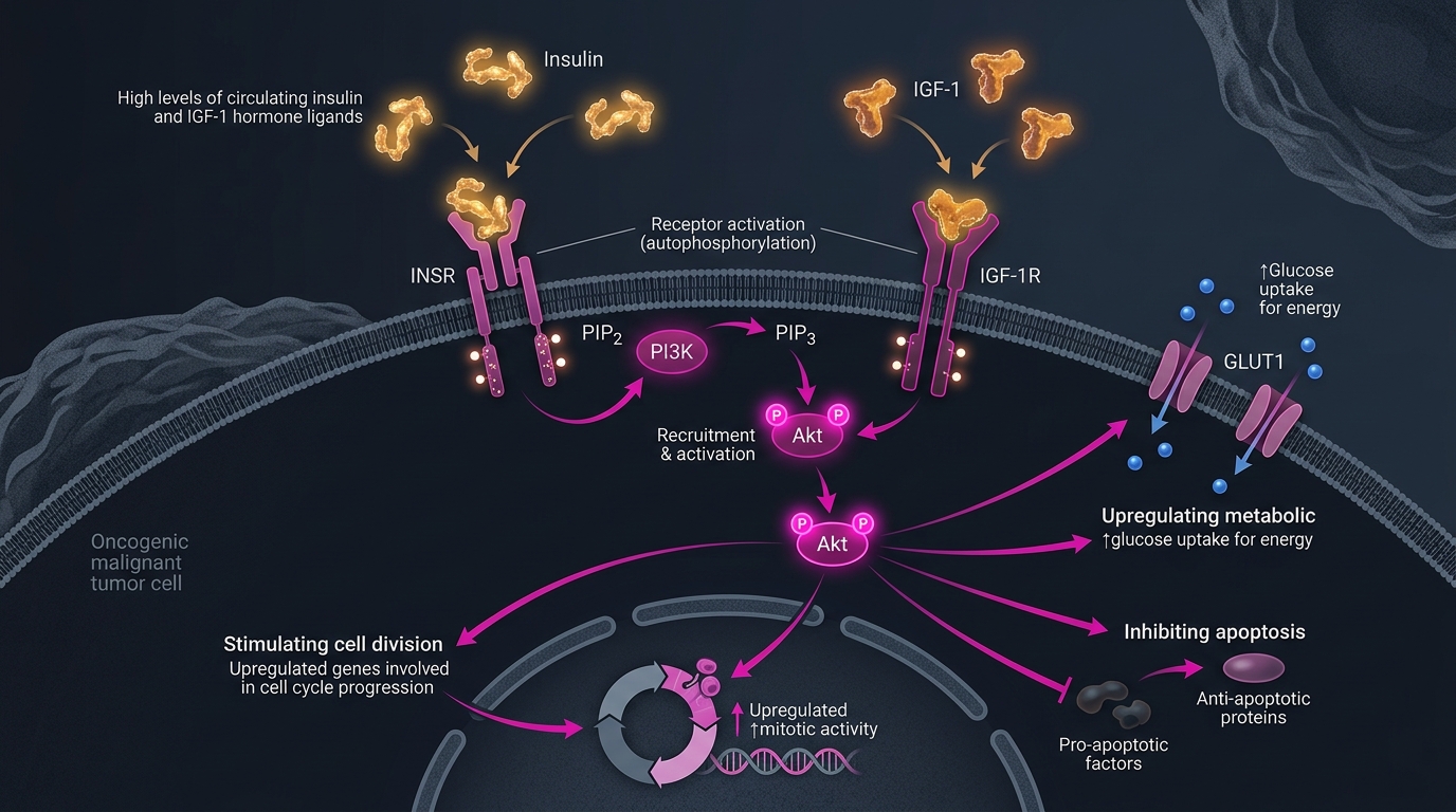
"If a genetic mutation represents the spark capable of igniting a brushfire, insulin and IGF-1 are the continuous blast of pure oxygen that fans it into an inferno. Under healthy physiological fasting, insulin drops, prompting cells to shift into recycling and debris maintenance (autophagy). But when insulin stays elevated day and night, the body never receives the biochemical signal to pause growth: the biological gas pedal is pinned to the floorboard without brakes."
1. The Molecular Cascade: From Membrane Receptor to Nuclear Mitosis
Many human malignancies (especially colorectal, breast, prostate, and endometrial carcinomas) overexpress insulin receptors (IR-A) and IGF-1 receptors (IGF-1R) at markedly elevated densities.
Upon ligand binding, insulin and IGF-1 activate the most notorious proliferative pathway in oncological biology:
Insulin / IGF-1 binds Tyrosine Kinase
PI3K Enzyme Activation
Akt Kinase Phosphorylation
mTORC1 Switch Triggered
Unchecked Mitosis & Apoptosis Arrest
- Igniting the PI3K - Akt Cascade: Receptor dimerization triggers autophosphorylation, recruiting insulin receptor substrates (IRS-1/2) and engaging phosphoinositide 3-kinase (PI3K). PI3K converts membrane to , creating a docking platform that recruits and fully activates Akt (Protein Kinase B).
- Engaging the mTORC1 Engine: Activated Akt phosphorylates and disables the TSC1/TSC2 tuberous sclerosis complex, releasing the small GTPase Rheb to powerfully stimulate mTORC1. In turn, mTORC1 phosphorylates S6K1 and 4E-BP1, accelerating ribosomal translation and biomass generation for mitosis.
- Silencing Apoptosis via FoxO Inactivation: Akt directly phosphorylates FoxO transcription factors, expelling them from the nucleus into the cytoplasm. Without nuclear FoxO, cells lose their ability to transcribe pro-apoptotic executioners like Bim and Bad. Defective cells that ought to undergo programmed cell death instead survive and replicate.
- Synergy with PTEN Loss: PTEN serves as the master phosphatase brake against PI3K signaling. In tumors harboring PTEN silencing mutations, hyperinsulinemia removes any remaining downstream restraint, locking the cell in a perpetual proliferative state.
2. Biological Comparison: Metabolic Equilibrium vs Chronic Hyperinsulinemia
| Biological Parameter | Healthy Metabolic Baseline | Chronic Hyperinsulinemia & Insulin Resistance |
|---|---|---|
| Fasting Serum Insulin () | Below 5 µIU/mL (Optimal) | Frequently above 15 - 25 µIU/mL |
| Hepatic IGFBP-1 & IGFBP-3 | Robust levels, sequestering free active IGF-1 | Suppressed by fatty liver, flooding serum with free bioactive IGF-1 |
| Intracellular mTORC1 Tone | Rhythmic, cycling smoothly between feeding and fasting | Continuously stimulated 24/7, suppressing protective autophagy |
| Programmed Apoptosis | Intact, systematically purging damaged cells | Strongly inhibited via Akt-mediated Bcl-2 survival upregulation |
| Pre-malignant Surveillance | Restrained by p53 and cell cycle checkpoints | Provided continuous growth signals and abundant substrate supply |
3. Three Clinical Levers to De-escalate the Insulin & IGF-1 Axis
Recognizing insulin's mitogenic role unlocks actionable non-toxic metabolic strategies:
- Intermittent Fasting & Breaking the Continuous Carbohydrate Wave: Every refined carbohydrate bolus prompts pancreatic beta-cells to secrete an insulin wave. Extending the inter-meal fasting window (such as a 16:8 protocol) permits basal insulin to decline to baseline, allowing AMPK to reactivate and dampen mTORC1.
- Stimulating Non-Insulin-Dependent Glucose Uptake via Skeletal Muscle: During muscle contraction in resistance training and brisk intervals, GLUT4 glucose transporters translocate to the sarcolemma via mechanical and AMPK-mediated pathways completely independent of insulin. This siphons off circulating glucose without placing demands on the pancreas.
- Reversing Visceral Adiposity & Restoring Tissue Sensitivity: Shedding ectopic fat within the liver and pancreas addresses the mechanical root of insulin resistance. Fermentable dietary fiber yields short-chain fatty acids (SCFAs), optimizing the gut-liver axis and restoring cellular insulin receptor sensitivity.
4. Key Clinical Takeaways
- Insulin and IGF-1 are potent mitogens: Beyond caloric clearing, they are primary upstream growth stimuli activating the PI3K-Akt-mTOR survival axis.
- Insulin resistance actively fuels tumor growth: Sustained hyperinsulinemia paired with suppressed IGFBP unbinds active IGF-1 to stimulate neoplastic clones.
- Lowering basal insulin is foundational prevention: Glycemic modulation, time-restricted eating, and muscular engagement serve as premier metabolic tools to lock down this proliferative switch.