
For decades, clinical practice guidelines recommended high-dose calcium supplementation for postmenopausal women and the elderly to prevent osteoporosis. However, recent large-scale clinical trials have unveiled a disturbing reality: calcium supplementation in isolation does not significantly reduce fracture rates but instead increases the risk of cardiovascular events by up to 30%. This medical phenomenon is known as the Calcium Paradox. Calcium, instead of entering the bone matrix, deposits in soft tissues, particularly the tunica intima of arteries, forming rigid atherosclerotic calcifications. To resolve this paradox, scientific research has turned to the synergistic axis of Vitamin D3 and Vitamin K2 (specifically MK-7). This combination is not merely additive; it is a mandatory, interdependent biochemical cascade at the molecular level designed to safely and effectively redistribute calcium within the human body.
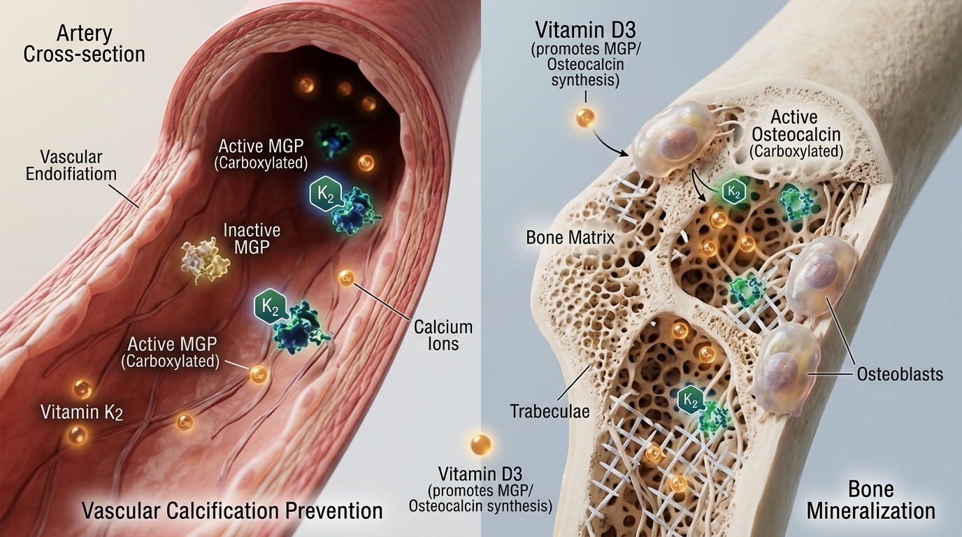
"Imagine Calcium as construction bricks, and Vitamin D3 as a fleet of trucks rapidly dumping these bricks into the construction site (the bloodstream). Without bricklayers to direct them, these bricks will pile up chaotically, blocking traffic and damaging the infrastructure (arterial calcification). Here, Osteocalcin and Matrix Gla Protein (MGP) are the bricklayers, but they are fast asleep (inactive). Vitamin K2 MK-7 acts as a strong cup of espresso, awakening these workers, enabling them to lay the bricks precisely into the building walls (bones) and sweep the debris off the highways (arteries)."
Molecular Pathway Flowchart
Vitamin D3 Ingestion
VDR Receptor Activation
Upregulation of inactive Osteocalcin & MGP (ucOC & ucMGP)
Vitamin K2 MK-7 activates Gamma-Glutamyl Carboxylase
Conversion to active cOC & cMGP
Directing Calcium to Bone & Inhibiting Arterial Calcification
1. Micro-Molecular Mechanisms: The Biochemical Interdependence of D3 and K2 MK-7
Upon ingestion, Vitamin D3 undergoes hydroxylation to become 1,25-dihydroxyvitamin D3 [1,25(OH)2D3], the biologically active ligand that binds to the intracellular Vitamin D Receptor (VDR). Activation of the VDR transcription factor upregulates the expression of key Vitamin K-dependent proteins (VKDPs): Osteocalcin (secreted by osteoblasts in bone tissue) and Matrix Gla Protein (MGP, synthesized by vascular smooth muscle cells [VSMCs] and chondrocytes). However, these newly synthesized proteins are initially in their inactive, uncarboxylated states, designated as ucOC and ucMGP, respectively. In this state, they lack the structural conformation required to bind divalent calcium ions (Ca2+).
To achieve functional activation, specific glutamic acid (Glu) residues on these proteins must undergo post-translational gamma-carboxylation to form gamma-carboxyglutamic acid (Gla) residues. This critical enzymatic reaction is catalyzed by Gamma-glutamyl carboxylase (GGCX), an enzyme embedded in the endoplasmic reticulum membrane. GGCX strictly requires the reduced form of Vitamin K2 (hydroquinone) as an obligate cofactor. During this reaction, Vitamin K2 is oxidized into Vitamin K epoxide, which is subsequently recycled back to its active form by the enzyme Vitamin K epoxide reductase (VKOR). In the absence of sufficient Vitamin K2, this recycling loop is compromised, leading to an accumulation of inactive ucOC and ucMGP. Consequently, free calcium ions circulate aberrantly and deposit within the arterial walls rather than being integrated into the hydroxyapatite crystal lattice of the bone matrix.
2. The Biological Paradox and Clinical Comparative Metrics
The biological paradox lies in the fact that high-dose Vitamin D3 supplementation in the state of Vitamin K2 deficiency actively exacerbates vascular calcification. Vitamin D3 enhances intestinal calcium absorption and drives the transcription of ucMGP. Without adequate Vitamin K2 to carboxylate ucMGP into its active form (cMGP), these inactive proteins cannot inhibit the nucleation of calcium phosphate crystals along the elastic fibers of the arterial wall. Conversely, active cMGP exhibits an exceptionally high affinity for calcium, acting as the most potent local inhibitor of vascular mineralization. Similarly, uncarboxylated osteocalcin (ucOC) cannot bind calcium to the bone scaffolding, leading to osteoporotic bones despite elevated systemic calcium levels.
| Physiological State | Protein Carboxylation Status | Clinical Endpoint |
|---|---|---|
| D3 and K2 Deficiency | Elevated ucOC, elevated ucMGP (Inactive) | Severe osteoporosis, high fracture risk, moderate arterial stiffness |
| Isolated D3 Supplementation (K2 Deficient) | Extremely high ucOC, extremely high ucMGP | Accelerated aortic calcification, increased atheromatous plaque burden, poor bone mineralization |
| Optimal D3 + K2 MK-7 Co-administration | Elevated cOC, elevated cMGP (Fully Active) | Enhanced bone mineral density (BMD), preserved arterial elasticity, minimized cardiovascular risk |
The comparative matrix clearly demonstrates that high-dose Vitamin D3 monotherapy can inadvertently trap the body in a calcium trap. Here, calcium is rapidly absorbed into systemic circulation but lacks molecular direction. The accumulation of circulating ucMGP serves as an independent prognostic biomarker for arterial stiffness and all-cause mortality in cardiovascular and chronic kidney disease (CKD) patients. Only when Vitamin K2 MK-7 is co-administered can the carboxylation process be completed, converting ucMGP into active cMGP, which actively decalcifies the arterial elastin fibers and restores vascular compliance.
3. Practical Application & Clinical Optimization Strategies
From a clinical perspective, optimizing the synergistic ratio and dosing of Vitamin D3 and K2 MK-7 is paramount to achieving the dual therapeutic benefit of bone preservation and cardiovascular protection. The MK-7 (Menaquinone-7) isoform of Vitamin K2 is highly preferred over MK-4 due to its longer isoprenoid side chain, which confers a prolonged biological half-life of approximately 72 hours (compared to just a few hours for MK-4). This extended kinetic profile allows MK-7 to maintain stable serum concentrations and achieve superior distribution to peripheral tissues, including the vascular wall and bone matrix.
Current clinical protocols suggest pairing a daily maintenance dose of Vitamin D3 (2000 IU to 5000 IU) with Vitamin K2 MK-7 (90 mcg to 180 mcg). For patients presenting with advanced arterial stiffness or severe osteoporosis, the therapeutic dose of MK-7 may be escalated up to 360 mcg daily under direct medical supervision. An essential safety guideline involves drug-nutrient interactions: patients on classic vitamin K antagonists (such as Warfarin or Coumadin) must consult their physicians before supplementing with K2, as it can directly counteract the anticoagulant efficacy. However, this interaction is not observed with novel oral anticoagulants (NOACs/DOACs) such as Rivaroxaban or Apixaban. For optimal bioavailability, these fat-soluble vitamins should always be administered alongside a meal containing healthy dietary lipids to maximize lymphatic absorption.