
Phytocodex emblemPhytocodex
🇬🇧 English Edition|Scientific Literature Synthesis
01 · A nanoscale hypodermic syringe piercing leukocyte walls
Type III Secretion Injectisome (T3SS)

Y. pestis docks against immune cells and deploys a hollow needle to pump cytotoxic effectors directly into the host cytosol, bypassing circulating antibodies.

02 · Molecular saboteurs freezing cellular defenses and alarms
The Cytotoxic Yop Arsenal (YopH, YopE, YopJ)

YopH/YopE depolymerize the actin cytoskeleton to paralyze phagocytosis, while YopJ silences NF-κB to prevent alarm cytokine release.

03 · A high-velocity respiratory strike with a 24-hour golden window
Primary Pneumonic Plague Aerosol

Inhaled bacteria replicate silently for 24 hours before unleashing fatal alveolar hemorrhage, requiring prompt antibiotic intervention within hours.

Clinical Deep-DiveImmune Literature-Synthesized & EBM-Verified 8 min read

The Phantom of Laboratory Plague: Deconstructing Yersinia pestis Type III Secretion Nanomachinery and Virulence Dynamics

Recent biosafety containment alarms at the Irkutsk Anti-Plague Research Institute in Siberia have renewed international scrutiny on high-consequence bacterial pathogens. Yersinia pestis is not merely the historical agent of the Black Death; it harbors an exquisitely engineered Type III Secretion System (T3SS) needle that directly translocates paralyzing Yop effectors into host immune cells, disarming macrophages and transforming alveolar beds into lethal hemorrhagic battlegrounds within 48 hours.

CH
Dr. Xuan Chien HoangDr. rer. nat. | University of Hamburg
2026-10-08T09:00:00ZDOI: 10.1016/j.tim.2015.11.008 3 Referenced Literature

In early October 2026, emergent reports concerning an unexplained fatality of a laboratory technician at the Irkutsk Anti-Plague Research Institute of Siberia and the Far East in Russia sparked profound global bio-surveillance scrutiny. While official statements attributed the tragedy to severe pneumonia of unknown etiology, the rapid medical quarantine of approximately two hundred personnel inevitably rekindled memories of the 1979 Sverdlovsk anthrax release and legacy military biological programs on Vozrozhdeniya Island. Among Tier-1 biological select agents, Yersinia pestis occupies a singular historical and clinical position. Having eliminated an estimated one-third of medieval Europe during the Black Death, the bacterium combines extreme lethality with aerosol transmissibility. What grants this Gram-negative coccobacillus the devastating capacity to dismantle human innate immunity within dozens of hours? The biochemical answer resides in its Type III Secretion System (T3SS), a molecular nanomachine functioning as a nanoscale hypodermic needle that perforates and neutralizes human immune defenses.
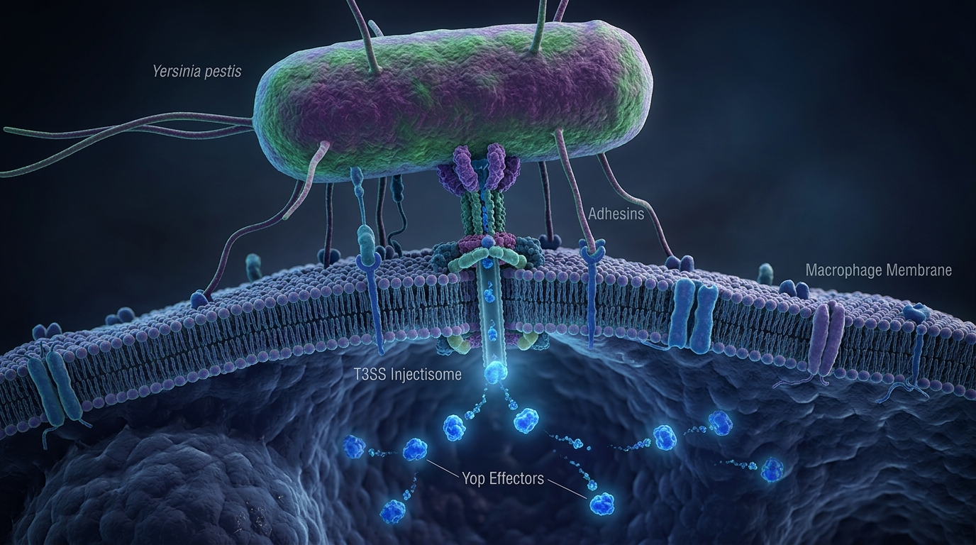
Molecular graphics of Yersinia pestis and the Type III Secretion System T3SS injectisome

"Picture a host macrophage as an armed biological fortress guarding peripheral tissues. Under standard physiological conditions, the instant an invading bacterium is detected, the fortress engulfs the pathogen inside phagosomes to dissolve it via acid and reactive oxygen species. Yersinia pestis, however, behaves like an infiltration unit wielding a nanoscale Type III syringe (T3SS). It docks against the fortress wall and injects paralyzing Yop toxins straight into the cellular command center. In seconds, defense artillery is disarmed, the macrophage cytoskeleton freezes, inflammatory sirens are silenced, and the fortress is transformed into a biological morgue."


Molecular Pathway Flowchart

Molecular Mechanism Pipeline
PHASE 01STIMULUS

Yersinia pestis Docks with Host Immune Cell

Next ➔Inspect
PHASE 02SIGNAL HUB

pCD1 Plasmid Activates T3SS Injectisome

Next ➔Inspect
PHASE 03SIGNAL HUB

Translocation of YopH, YopE, YopJ Effectors

Next ➔Inspect
PHASE 04INHIBITION

Inhibition of MAPK/NF-κB & Actin Depolymerization

Next ➔Inspect
PHASE 05INHIBITION

Paralysis of Phagocytosis & Suppression of Alarm Cytokines

Next ➔Inspect
PHASE 06ENDPOINT

Uncontrolled Bacterial Expansion in Blood & Lungs

Terminal ✓Inspect

1. Nanoscale Architecture of the Type III Secretion Injectisome

Unlike enteric ancestors such as Yersinia pseudotuberculosis, Yersinia pestis achieved evolutionary hypervirulence primarily through horizontal acquisition of distinct plasmids, foremost among them the 70-kilobase pCD1 virulence plasmid. This plasmid encodes the multiprotein Type III Secretion System (T3SS), commonly designated the injectisome.

Triggered upon sensing mammalian host temperature (37 degrees Celsius) and low extracellular calcium concentrations:

  • The Basal Body: Spans both the bacterial inner and outer membranes as well as the peptidoglycan wall, driven by the YscN ATPase engine at the cytoplasmic face.
  • The Needle Filament: Assembled via helical polymerization of hundreds of YscF subunits, projecting roughly 40 nanometers beyond the lipopolysaccharide capsule with an inner hollow lumen diameter of barely 2 nanometers.
  • The Translocon Complex: Comprising YopB and YopD proteins. Upon direct physical adhesion to target leukocytes, YopB and YopD insert into the eukaryotic plasma membrane to form a continuous pore, allowing cytotoxic effector proteins to travel straight from the bacterial cytosol into host cytoplasm without systemic antibody exposure.

2. The Cytotoxic Yop Arsenal: Molecular Disarmament of Innate Immunity

Once the translocon conduit is established, Yersinia pestis orchestrates a simultaneous biochemical assault on intracellular signaling nodes using Yop (Yersinia outer proteins) effectors:

  1. YopH (Protein Tyrosine Phosphatase): Regarded as one of the most catalytically potent phosphatases discovered in nature. YopH selectively dephosphorylates focal adhesion proteins, including p130Cas and FAK, severing the signaling cascades requisite for phagocytic cup formation. The macrophage becomes physically incapable of extending pseudopodia.
  2. YopE and YopT (Rho GTPase Disrupters): Function as GTPase-activating proteins (GAPs) and cysteine proteases that target small Rho-family GTPases (RhoA, Rac1, Cdc42). By locking these molecular switches in an inactive state, YopE triggers swift depolymerization of the actin cytoskeleton, causing target immune cells to round up and detach.
  3. YopJ (Serine/Threonine/Lysine Acetyltransferase): The master suppressor of immune alarm systems. YopJ acetylates key kinase residues in the MAPK and IKK signaling complexes, irreversibly preventing NF-kappa-B activation. Consequently, transcription of vital pro-inflammatory alarm cytokines (TNF-alpha, IL-1beta) is silenced, while rapid leukocyte apoptosis is triggered.

Below is a clinical and molecular comparison highlighting the divergence between routine bacterial encounters and the paralysis induced by Yersinia pestis:

Biomarker & Clinical MetricConventional Infection (E. coli, S. aureus)Yersinia pestis T3SS Pathogenesis
Leukocyte Phagocytic CapacityRobust, prompt engulfment and lysosomal acidificationCompletely paralyzed via YopH/YopE actin disruption
Pro-inflammatory Cytokine ResponseRapid surge of TNF-alpha, IL-6 rallying reinforcementsSilenced during early incubation by YopJ NF-kappa-B arrest
Bacterial Proliferation KineticsControlled and localized by local tissue barriersExponential systemic expansion in lymph nodes and blood
Primary Clinical PresentationLocal abscess, controlled fever, tissue erythemaNecrotic Buboes (Bubonic) or Fatal Pulmonary Hemorrhage
Therapeutic Window for Antibiotics3 to 5 days from onsetExtremely narrow: Must initiate within the first 24 hours

3. Biosafety Guidelines & Practical Clinical Protocols for Pneumonic Plague

While classic bubonic plague arises following the bite of an infected flea vector (Xenopsylla cheopis), respiratory inhalation of aerosolized droplets generates primary pneumonic plague. This is the exact clinical manifestation feared in laboratory exposure incidents or biosecurity breaches.

The pathophysiology of primary pneumonic plague unfolds with terrifying velocity:

  1. The Biphasic Immune Stealth Window (0 to 24 hours): Because T3SS-delivered YopJ suppresses alveolar inflammatory signaling, bacteria multiply unchecked within pulmonary parenchyma without eliciting early respiratory distress. The patient appears clinically stable while trillions of organisms colonize alveolar airspaces.
  2. The Pro-inflammatory Hyper-reaction (24 to 48 hours): Once bacterial thresholds exceed critical mass, overwhelming lysis of alveolar-capillary membranes triggers a delayed, catastrophic cytokine storm. Patients develop high fever, acute dyspnea, and produce watery, blood-tinged sputum teeming with viable bacillary chains. Rapid destruction of the pulmonary architecture culminates in fatal acute respiratory distress syndrome (ARDS) and hemodynamic collapse.
  3. Direct Human-to-Human Aerosol Transmission: Unlike bubonic cases which require arthropod vectors, pneumonic plague transmits directly via respiratory droplets produced during coughing. In confined facilities lacking negative-pressure BSL-3/BSL-4 engineering controls, secondary infection rates increase exponentially.

Practical Protocols and Antimicrobial Guidelines:

Modern clinical management requires immediate administration of parenteral aminoglycosides (gentamicin, streptomycin) or fluoroquinolones (ciprofloxacin, levofloxacin) within the first 24 hours of fever onset. Once the infection passes into late pneumonic necrosis, bactericidal killing releases massive amounts of endotoxin, leaving mortality rates above 90% even with aggressive intensive care. Biosafety transparency, stringent containment protocols, and vigilant molecular diagnostics remain the indispensable barricades protecting civilization from the return of its oldest microbial nemesis.

§ 6 · References & Primary Evidence

Verified Source List.

References indexed via NCBI PubMed & CrossRef

  1. 01
    The Yersinia Ysc-Yop 'type III' weaponry

    Nature Reviews Molecular Cell Biology · 2002

    Pathogenic Yersinia species disarm host immune defenses by using a type III secretion apparatus to inject Yop effectors directly into the host cell cytosol, paralyzing macrophages and blocking phagocytosis.

    Landmark Molecular Reviewn = systematic/in-vitro
  2. 02
    Pneumonic Plague: The Darker Side of Yersinia pestis

    Trends in Microbiology · 2016

    Comprehensive analysis of primary pneumonic plague pathogenesis: the biphasic course from early anti-inflammatory stealth phase to catastrophic alveolar necrosis, cytokine storm, and fatal hemorrhagic destruction.

    Pulmonary Pathophysiological & Virulence Reviewn = systematic/in-vitro
  3. 03
    The Sverdlovsk anthrax outbreak of 1979

    Science · 1994

    Independent genetic and epidemiological verification of accidental aerosol release from a high-containment microbiology facility, establishing foundational global standards for bio-risk oversight and aerosol containment.

    Epidemiological and Forensic Biological Containment Investigationn = systematic/in-vitro
Interactive Laboratory Simulation

Model calculations execute entirely client-side.

Gizmo 02Bản đồ Con đường Tín hiệu & Thụ thể Tế bào
kịch bản mẫu

Điều khiển tham số

Thư viện hoạt chất

Thụ thể tiếp nhận

Tiểu đơn vị IKKβ / NF-κB p65

0 byte truyền ngoài

MÀNG TẾ BÀO (PLASMA MEMBRANE)Curcumin (Tinh chất Nghệ)CUCurcuminSulforaphane (Mầm Súp Lơ)SUSulforaphaneBerberine (Cây Hoàng Liên)BEBerberineQuercetin (Vỏ Táo & Hành Tây)QUQuercetinNF-κB THỤ THỂ↓Giảm phosphory↓Ngăn thoái giá↓Chặn chuyển vị↓Giảm các chất NHÂN TẾ BÀOADN → mARNSƠ ĐỒ MÔ PHỎNG — KHÔNG THEO TỶ LỆ TUYỆT ĐỐI

Đích tác động

NF-κB

Nút hạ nguồn

4vị trí

Chiều tác động

GIẢMđiều hòa

Khoang tế bào

BÀO TƯƠNG→NHÂN

Phân tích khoa học

Curcumin (Tinh chất Nghệ) → Các phân tử curcuminoids làm ngắt quãng dòng tín hiệu viêm kinh điển NF-κB ngay tại nút IKKβ, ngăn chặn giải phóng phức hợp gây viêm.Chuỗi tác động hạ nguồn: ↓ Giảm phosphoryl hóa IKKβ · ↓ Ngăn thoái giáng IκBα · ↓ Chặn chuyển vị p65 vào nhân · ↓ Giảm các chất viêm TNF-α, IL-6, COX-2

💡 Góc Giải Thích Y Khoa Dễ Hiểu

Hiểu sâu bản chất sinh hóa của tế bào qua những hình tượng ẩn dụ thực tế:

NF-κB NODE
Cắt đứt dây chuông báo động viêm nhiễm NF-κB

NF-κB giống như chiếc 'chuông báo cháy' của tế bào. Khi bị tổn thương, stress oxy hóa hoặc vi khuẩn kích thích, chiếc chuông này reo liên hồi gây sưng đau, viêm khớp và viêm mạn tính. Curcumin đi thẳng vào nút IKKβ và 'ngắt dây chuông', dập tắt tín hiệu viêm ngay từ tế bào chất trước khi nó kịp kích hoạt nhân tế bào sản sinh độc tố viêm.

Ý nghĩa Lâm sàng Thực tế

Giảm sưng đau khớp, hạ chỉ số viêm hs-CRP và bảo vệ niêm mạc ruột.

Nguồn chiết xuất: Củ nghệ vàng (Curcuma longa)
Share this Dispatch
CH

Dr. Xuan Chien Hoang

Doctor of Natural Sciences (Univ. of Hamburg) · Founder, Lava Health GmbH

Biomedical scientist and product developer with over a decade of international experience in Germany and APAC. Author of The Cancer Code (Amazon: eBook, Paperback, Hardcover). Pioneering the East-West botanical bridge, bioavailability enhancement, and data-driven HealthTech.

PHYTOCODEX · WEEKLY BRIEF

Evidence-Based Biomedical Intelligence Straight to Your Inbox

Deep-dives into bioavailability kinetics, cellular pathways, interactive simulation models, and East-West ethnobotanicals by Dr. Xuan Chien Hoang. Zero spam, unsubscribe anytime.

GDPR compliant. 1-click unsubscribe anytime.
Related Analytical Dispatches
Cellular Aging

Urolithin A từ Quả Lựu: Cơ chế Kích hoạt Mitophagy Dọn dẹp Ty thể Độc lập với NAD+ Giúp Phục hồi Sức bền Cơ bắp

Nhiều người tin rằng chỉ cần uống nước ép lựu là đủ để có được các lợi ích trẻ hóa tế bào của Urolithin A. Tuy nhiên, sự thật sinh học phức tạp hơn nhiều: cơ thể chúng ta không tự sản sinh ra Urolithin A. Nó là sản phẩm chuyển hóa thứ cấp của hệ vi sinh đường ruột từ ellagitannins có trong lựu, và chỉ có khoảng 30 đến 40 phần trăm dân số sở hữu hệ vi sinh phù hợp để thực hiện quá trình chuyển đổi này. Bài viết bóc tách cơ chế phân tử độc đáo của Urolithin A trong việc kích hoạt mitophagy (quá trình tự thực ty thể) thông qua con đường độc lập với NAD+, giúp dọn dẹp các ty thể già cỗi, phục hồi hiệu suất cơ bắp mà không làm cạn kiệt nguồn dự trữ năng lượng nội bào.

Open Dispatch
Cellular Aging

Urolithin A from Pomegranate: Deciphering the NAD+ Independent Mitophagy Pathway for Muscle Recovery and Mitochondrial Longevity

While pomegranate is celebrated as an anti-aging superfood, drinking its juice rarely yields the therapeutic levels of its active metabolite, Urolithin A. This molecule is not directly present in pomegranates: instead, it requires specific gut microbiota to convert dietary ellagitannins into Urolithin A. Crucially, only 30 to 40 percent of humans possess the microbial profile capable of this conversion, leaving the majority as non-responders. This monograph dissects the molecular mechanism of Urolithin A as a first-in-class mitophagy activator that operates independently of the classic NAD+ pathway, bypassing cellular energy depletion to selectively clear damaged mitochondria, enhance ATP synthesis, and restore skeletal muscle function in aging populations.

Open Dispatch
Immune

Bóng Ma Dịch Hạch Từ Phòng Thí Nghiệm Nga: Giải Mã Cỗ Máy Tiêm Độc T3SS và Độc Lực Của Vi Khuẩn Yersinia pestis

Vụ việc rò rỉ mầm bệnh tại Viện Nghiên cứu Chống Dịch hạch Irkutsk (Nga) đặt giới y học toàn cầu vào tình trạng báo động đỏ. Yersinia pestis không chỉ là tác nhân gây nên thảm họa Cái Chết Đen trong lịch sử, mà còn sở hữu cỗ máy bơm độc lực Type III (T3SS) có khả năng tiêm thẳng protein độc tố Yop vào tế bào miễn dịch, làm tê liệt đại thực bào và biến lá phổi thành bãi chiến trường xuất huyết tử vong chỉ sau 48 giờ.

Open Dispatch
The Phantom of Laboratory Plague: Deconstructing Yersinia pestis Type III Secretion Nanomachinery and Virulence Dynamics · Phytocodex · Phytocodex